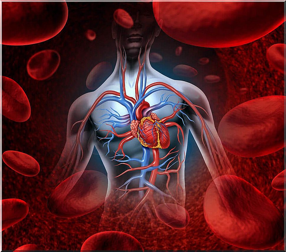
The Heart, The Body’s Pump
It always beats, but we realize it when we exercise, when we see the person we like and when we get scared. We know that it helps the body to stay alive and that is why it is called the “body pump.” In effect, we are referring to the heart, the organ that pumps blood to the rest of the body.
Below we will share some common questions and answers that allow you to better understand the functioning of this vital organ.
Where is the heart located?
It is located in the thorax, in the region known as the “middle mediastinum”. It is located to the left of the midline, which explains why this lung is smaller. The anterior aspect of the heart lies behind the sternum and thymus.
The lateral faces border the lungs, while the vertex, the lower part, rests on the diaphragm. The most posterior part of the heart lies in front of the descending aorta.
How is the heart formed?
The heart is made up of four chambers: two atria and two ventricles. We can divide it into “left heart” and “right heart”.
It should be noted that the cavities, on each side, are communicated by a system of valves that prevent the backward flow of blood. Between both sides of the heart, right and left, is the septum or septum. This is in turn divided into two parts:
- Interatrial septum, separating the two atria.
- Interventricular septum, separating the two ventricles. This part of the septum is of utmost importance. In it is the nerve network that transmits electrical impulses, maintaining the connection between atria and ventricles.
On the right
The right atrium receives blood from the entire body through two large veins: the cavae. The superior vena cava collects blood drainage from the upper half of the body.
The inferior vena cava carries blood from the lower half to the heart. In addition, it also receives blood from the coronary system, which supplies the heart itself.
The right atrium is separated from the right ventricle by the tricuspid valve. It is made up of three leaflets that close when the ventricle contracts, preventing the backward movement of blood.
The pulmonary arteries start from the right ventricle, in charge of carrying blood to the lungs for oxygenation. Between the ventricle and the outlet of the vessels is another valve system: the pulmonary valve. It opens with each ventricular contraction, allowing blood to flow out, and closes just after, preventing its backward movement.
Read: Blood types
On the left
Oxygenated blood from the lungs reaches the left atrium through the pulmonary veins. This chamber communicates with the left ventricle through the mitral (bicuspid) valve, made up of two leaflets.
The aorta, the great artery from which all the other arteries of the body will originate, starts from the left ventricle. As with the pulmonary arteries, there is a valve, the aortic, which prevents the backward movement of blood.
To understand the functioning of the heart even better, you can consult this link about the cardiac cycle.
What are the layers of the heart like?
- Endocardium : it is the innermost layer of the heart. It covers the interior of the cavities and continues with the endothelium of the blood vessels.
- Myocardium: It is the middle layer, the muscular layer. It is made up of muscle cells with different characteristics from other muscle fibers, cardiomyocytes. In this layer are the cells that produce atrial natriuretic peptide (ANP). It is an important vasodilator that works by lowering blood pressure. In addition, in this layer is the electrical system of the heart.
- Pericardium : it is the outermost layer of the heart, fibrous in nature. It forms a protective “sac” that surrounds the heart and continues until the exit of the great vessels.
Discover: Heart rate: what is it and how is it measured?
What is the irrigation and innervation of the heart?
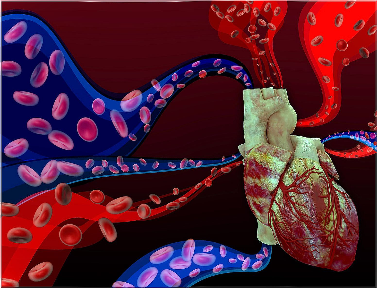
The heart has its own blood supply system: the left and right coronary arteries. They are the first branches that the aorta emits as soon as it leaves the left ventricle. In turn, each of them branches into a series of small vessels.
Venous drainage is done through the coronary veins. They converge into each other until they flow into the coronary sinus, the venous structure that carries blood to the right atrium. This ensures that the heart has all the oxygen it needs.
Although the heartbeat depends on the cardiac excitoconduction system itself, the organ is subject to autonomous regulation. That is, although the beats originate in the heart itself, its regulation depends on the sympathetic and parasympathetic systems.
The vagus nerve provides parasympathetic fibers. In general, its stimulation reduces the heart rate.
The sympathetic fibers are carried by fibers from the cervical sympathetic ganglia. In general, its stimulation increases the heart rate.
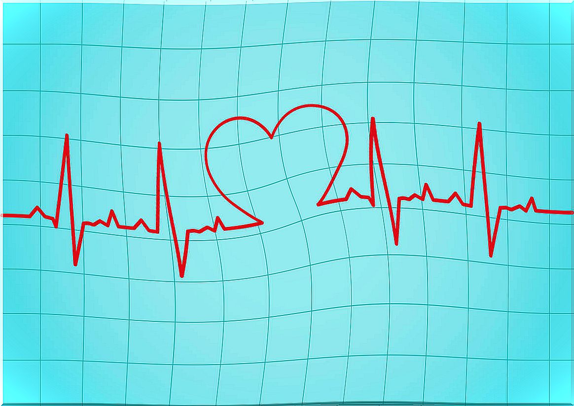
How does the heart beat?
The beats are born in the sinus node, the “pacemaker of the heart.” It is a small mass of modified muscle cells, specialized in the generation and transport of electrical impulses. It is like a small button located at the mouth of the superior vena cava in the right atrium.
An electrical impulse runs through the atria, causing their synchronized contraction during cardiac systole. Thus, the blood passes from the atria to the ventricles, which are relaxed.
In the interventricular septum there are two structures of special importance: the AV node and the bundle of Hiss. The AV node is a structure similar to the sinus node, capable of generating beats, albeit at a lower rate.
The Hiss bundle is a muscular cord, specialized in the transmission of the electrical impulse. It is “the wiring of the ventricles.”
When the electrical impulse reaches the ventricles, they contract, expelling blood into the pulmonary and peripheral circulation. We speak of systole when the ventricles contract and expel blood. By diastole, we mean ventricular relaxation that allows it to fill.
What curiosities about the heart do you have to know?

- Fetal circulation. During intrauterine life, the atria remain communicated in the absence of pulmonary respiration through the foramen ovale. With the first breaths outside the uterus, a pressure set is established that closes it. The extrauterine circulation is thus established.
- Fetal heartbeat: The first heartbeats of a fetus occur around day 21.
- Situs inversus. As a simple curiosity. This is an extremely rare anomaly. In it, either all the organs, or just a few, appear turned sideways, as in a mirror. That is, these patients have their heart on the right side. If they are listened to normally, no heartbeat is detected.
Did you know all this about the human heart? Whether the answer is yes or no, one thing is clear: the human body is incredible.