Anatomy Of The Skin
Have you ever been told that the skin is the largest organ in the body and that it has a complex structure, but also fascinating? Are you curious to know more about it so that you can start providing more appropriate care? If so, let’s get right to it!
The skin and its accessory structures make up the so-called integumentary system, which provides the body with general protection. And as we mentioned earlier, it is made up of multiple layers of cells and tissues.
The deepest layer of the skin is well vascularized (that is, it has numerous blood vessels). It also has numerous sensory, autonomous, and sympathetic nerve fibers that ensure communication to and from the brain.
To understand more about its structure, below we will divide it into parts and comment on each of them.
Layers of skin
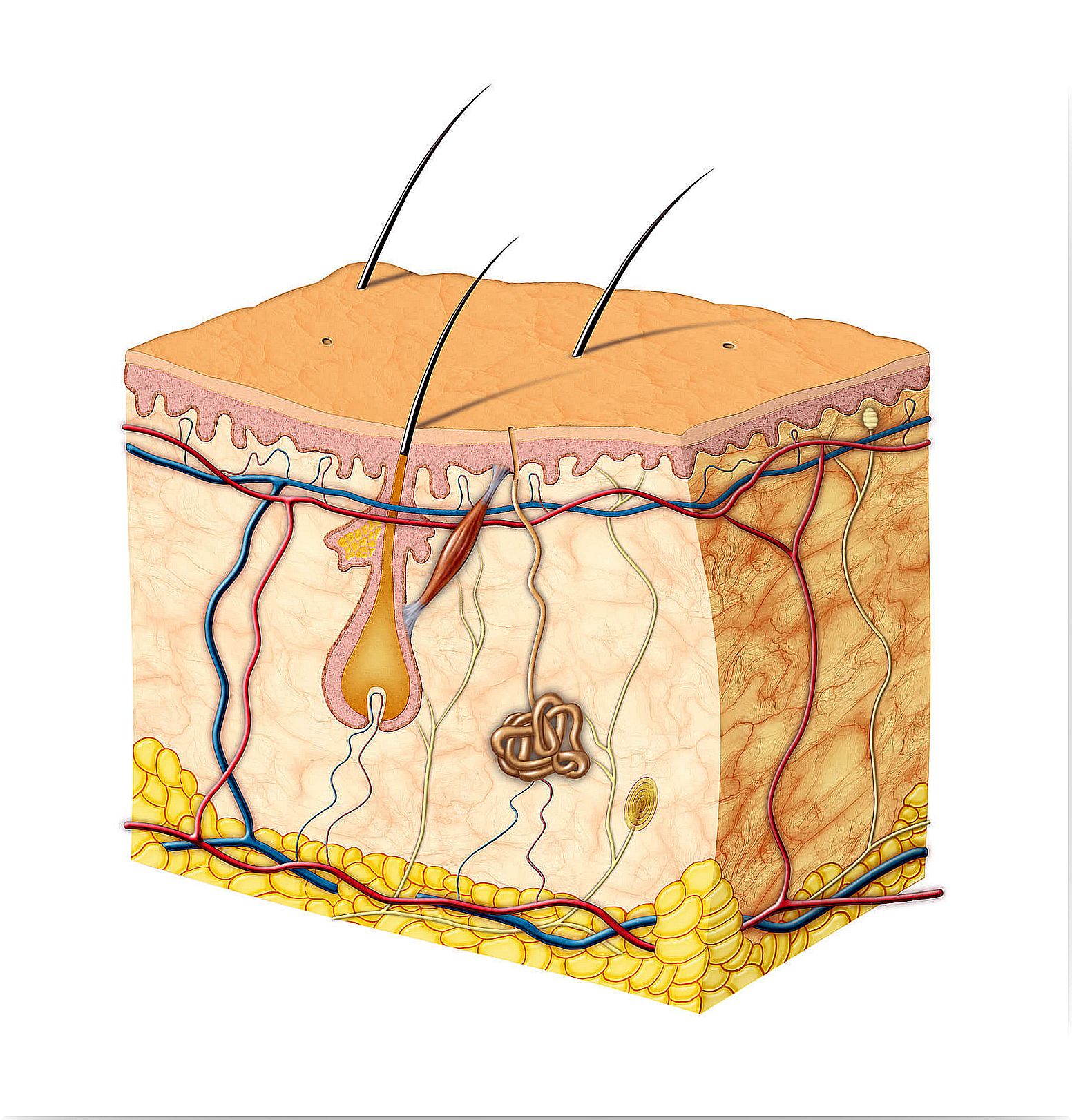
Basically, the skin is made up of two layers that cover a third layer of fat. These three layers differ in function, thickness, and strength.
Epidermis
The epidermis is the outermost layer and protects the body from the environment. It contains melanocytes (the cells in which melanoma develops), Langerhans cells (which are involved in the skin’s immune system), Merkel cells, and sensory nerves.
- Its thickness varies according to the type of skin.
- Only 0.05 millimeters thick on the eyelids and 1.5 millimeters on the palms of the hands and soles of the feet.
In turn, the epidermis is made up of five sub-layers that work together to rebuild the skin’s surface.
The basal cell layer
- The basal cell layer is the innermost layer of the epidermis and contains small round cells called basal cells.
- Basal cells continually divide and new cells push older cells to the surface, where they eventually shed.
- The basal cell layer is also known as the germinal stratum due to the fact that it is constantly germinating (producing) new cells.
- This layer contains cells called melanocytes that produce the skin pigment known as melanin , which gives it its brown color and helps protect the deeper layers from the harmful effects of the sun.
- Merkel cells, which are touch cells of neuroectodermal origin, are also found in the basal layer of the epidermis.
The squamous cell layer
- The squamous cell layer is located above the basal layer, and is also known as the stratum spinosum because the cells are held together with spiny projections.
- Within it are basal cells that have been pushed up, however these maturing cells are now called squamous cells or keratinocytes.
- Keratinocytes produce keratin , a tough, protective protein that makes up most of the structure of skin, hair, and nails.
- The squamous cell layer is the thickest layer of the epidermis and is involved in the transfer of certain substances in and out of the body.
- The squamous cell layer also contains so-called Langerhans cells. These attach to antigens that invade damaged skin and alert the immune system to their presence.
The stratum granular and the lucid stratum
- The keratinocytes of the squamous layer are pushed up through two thin epidermal layers called the stratum granulosa and the stratum lucid.
- As these cells move further to the surface of the skin, they get larger and flatter, stick together, and eventually dehydrate and die.
- This process results in the cells coalescing into layers of strong and durable material, which continue to migrate to the surface of the skin.
The stratum corneum
- The stratum corneum is the outermost layer of the epidermis, and is made up of 10 to 30 thin layers of dead keratinocytes, continuously lost.
- It is also known as the horny layer because its cells harden like an animal’s horn.
- As the outermost cells age and wear down, they are replaced by new layers of strong, long-lasting cells.
- The stratum corneum is continually shed as new cells take its place, but this shedding process slows down with age.
- Complete cell renewal occurs every 28 to 30 days in young adults, while the same process takes 45 to 50 days in older adults.
Dermis
The dermis lies below the epidermis and is the thickest of the three layers of skin (1.5 to 4 millimeters thick), making up about 90% of the skin’s thickness.
- The main functions of the dermis are to regulate temperature and supply the epidermis with nutrient-saturated blood.
- Much of the body’s water supply is stored within the dermis. This layer contains most of the specialized cells and structures that include blood vessels, lymphatic vessels, hair follicles, sweat glands, sebaceous glands, nerve ends, collagen, and elastin.
Blood vessels
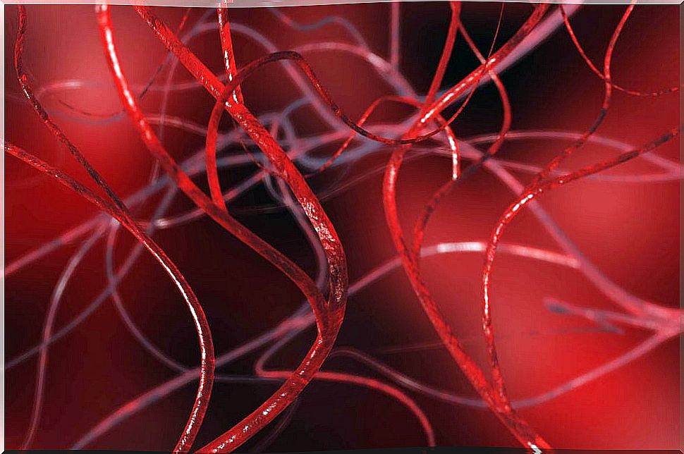
- Blood vessels supply nutrients and oxygen to the skin and remove cellular debris and cellular products.
- Blood vessels also carry vitamin D made in the skin back to the rest of the body.
Lymphatic vessels
- Lymphatic vessels bathe skin tissues with lymph, a milky substance that contains cells of the immune system to fight infection.
- These cells work to destroy any infection or invasion of organisms as the lymph circulates to the lymph nodes.
Hair follicles
A hair follicle is a tubular sheath that surrounds the part of the hair that is under the skin and nourishes it.
Sweat glands
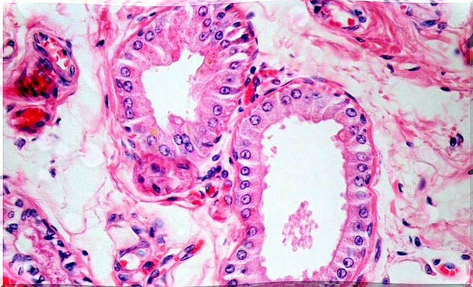
The average person has around three million sweat glands. These are classified according to two types:
-
Apocrine glands – These are specialized sweat glands that can be found only in the armpits and pubic region. These glands secrete a milky sweat that stimulates the growth of the bacteria responsible for body odor.
-
Eccrine glands: they are the true sweat glands. These glands, which are found throughout the body. They regulate body temperature by bringing water through the pores to the surface. There it evaporates and reduces the temperature. They can produce up to two liters of sweat per hour. However, they primarily secrete water, which does not encourage the growth of odor-producing bacteria.
- Sebaceous glands: These are attached to the hair follicles and can be found everywhere on the body except the palms of the hands and the soles of the feet. These glands secrete oil that helps keep the skin soft and supple. It also helps keep skin waterproof and protects against bacterial and fungal overgrowth.
Nervous ends
- The dermis layer also contains pain and tactile receptors that transmit sensations of pain, itching, pressure, and information regarding temperature in the brain for interpretation.
- If necessary, the tremor (involuntary contraction and relaxation of the muscles) is activated, generating body heat.
Collagen and elastin
- The dermis is held together by a protein called collagen, produced by fibroblasts.
- Fibroblasts are cells that give skin its strength and resistance.
- Collagen is a tough, insoluble protein found throughout the body in connective tissues that hold muscles and organs in place.
- In the skin, collagen supports the epidermis, which gives it its durability.
- The elastin , a similar protein, the substance that allows the back skin to its site when stretched and remains flexible.
The dermis is made up of two sublayers:
-
Papillary layer or superior; It contains a thin arrangement of collagen fibers. Provides nutrients for the layers of the epidermis and regulates temperature. Both of these functions are accomplished with a thin, extensive vascular system that functions similarly to other vascular systems in the body. With constriction and expansion they control the amount of blood that flows through the skin. This dictates whether body heat is dissipated when the skin is hot or is retained when it is cold.
-
Reticular or lower layer: it is made of thick collagen fibers arranged parallel to the surface of the skin. It is denser than the papillary dermis and strengthens the skin, providing structure and elasticity. It is also compatible with other components, such as hair follicles, sweat glands, and sebaceous glands.
The subcutis
The subcutis is the innermost layer of the skin and consists of a network of fat and collagen cells. It is also known as the hypodermis or subcutaneous layer. It works as an insulator that conserves the body’s heat and, at the same time, as a shock absorber since it protects the internal organs. It also stores fat as an energy reserve for the body.
Blood vessels, nerves, lymphatic vessels, and hair follicles also pass through this layer. The thickness of the subcutaneous layer varies throughout the body and from person to person.
In short, the skin collects sensory information from the environment. It also plays a fundamental role in protecting the immune system and regulating temperature. In addition, it is capable of storing water, fat and vitamin D.
All this is possible thanks to the fact that it is made up of several fabrics that work together as a single structure.
The skin is a complex and fascinating structure
The skin is made up of different layers that fulfill specific functions in the body. We can consider it a complex structure, since it goes beyond what we can see externally.
On the other hand, it has very important functions both at a sensory level and in relation to the immune system. It is even key to storing fat and synthesizing important nutrients such as vitamin D. Did you know?