Physiology Of The Skin
The physiology of the skin refers to the largest organ of the body: the skin. The skin constitutes an important portion of the integumentary system, which also consists of accessory structures such as hair, nails, sweat, and sebaceous glands.
Together, the components of the integumentary system provide a first line of defense, whether against chemical or microorganism attack, physical impact or abrasion. This system also provides the sensory structures for sensing touch, pressure, pain, and temperature.
Anatomy and physiology of the skin: main layers
Epidermis
The epidermis is the outermost layer of the skin. It is composed mainly of keratinocytes, melanocytes, Langerhans cells, Merkel cells, and sensory nerves.
Therefore, the epidermis is made up of five sub-layers that work together to continually rebuild the surface of the skin. Thus, its parts are: the basal cell layer, the squamous cell layer, the stratum granulosa, the stratum lucid and the corneum.
Dermis
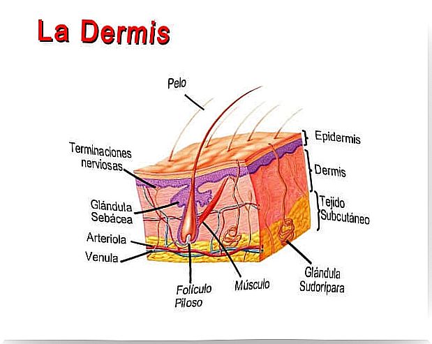 The dermis lies below the epidermis and is the thickest of the three.
Its main functions are to regulate temperature and supply nutrients to the epidermis. This is because the epidermis lacks blood capillaries so it depends on the blood supply to the dermis. The thermoregulatory function is due to the fact that said irrigation can contract by vasoconstriction in cold weather and expand by vasodilation in hot weather.
Much of the body’s water supply is stored within the dermis. It contains the blood and lymphatic vessels, hair follicles, sweat glands, and sebaceous glands, as well as nerve endings, collagen, and elastin. On the other hand, the dermis is made up of two sublayers: papillary and reticular.
Hypodermis
The hypodermis is the innermost layer of the skin and consists of a network of fat and collagen cells.
It works as an insulator, conserving body heat, and as a shock absorber protecting internal organs. It also stores fat as an energy reserve for the body.
Blood vessels, nerves, lymphatic vessels, and hair follicles also pass through this layer. The thickness of the subcutaneous layer varies throughout the body and from person to person.
Structures of the integumentary system
The integumentary system includes the cutaneous membrane (skin), its organs, structures and accessories. Therefore, this is the coating that acts as a protective and waterproof. It is self-healing, insulating, padded and stretchy.
The integumentary system contains most of the specialized cells and structures including:
Blood vessels
Blood vessels carry nutrients and oxygen-rich blood to the cells that make up the layers of the skin. In addition, waste products are taken away.
E l pili muscle arrector
The arrector pili is a small muscle in the physiology of the skin that is connected to each hair follicle and to the skin. When it contracts, it causes the hair to stand up and form “goose bumps.” Its function appears to be to provide insulation, as air is trapped between the upright hair.
Hair follicle
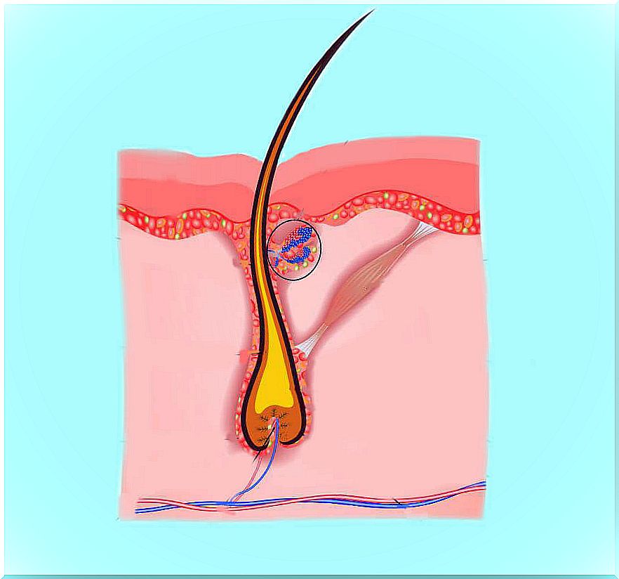
The hair follicle is a tube-shaped envelope that surrounds the part of the hair that is under the skin and nourishes it. It is found in the epidermis and dermis. It is the part of the skin that gives hair growth by concentrating stem cells, forming from a tubular invagination. Each hair rests on a hair follicle, this being the most dynamic skin structure and one of the most active in the entire body.
Langerhans cells
Langerhans cells bind to antigens that invade damaged skin and alert the immune system to their presence. They are abundant in the epidermis.
Melanocyte
A melanocyte is a cell that produces melanin and is found in the basal layer of the epidermis. Melanocytes are responsible for the pigment in the skin – known as melanin – that gives the skin its brown color and helps protect it from the harmful effects of the sun.
Sun exposure causes melanocytes to increase melanin production to protect the skin from harmful ultraviolet rays. Similarly, melanin patches on the skin cause birthmarks, freckles, and age spots.
Merkel cells
Merkel cells are a special type of cell found directly under the epidermis. These cells are very close to the nerve endings that receive the sensation of touch and can participate in the sense of touch. Cells also contain substances that can act like hormones.
Pacini corpuscle
The corpuscle of Pacini is a nerve receptor that responds to pressure and vibration. They are found mainly in the subcutaneous connective tissue and in the reticular dermis and are especially numerous in the hands and feet.
Sebaceous glands
The sebaceous glands are found in the physiology of the skin, especially in the dermis. Its main mission is to generate sebum that is responsible for protecting against microbes, thanks to the natural acidity of sebum, and lubricating the hair.
Sensory nerves
The physiology of the skin also includes sensory nerves. These nerves sense and transmit heat, pain, and other sensations. When something is not working properly and a skin biopsy is performed, the swelling, diameter, branching, contiguity, and general health of the sensory nerves are evaluated.
Stratum corneum
This is the outermost layer of the epidermis and is made up of dead skin cells. Protects the living cells beneath it by providing a tough barrier between the environment and the lower layers.
Sweat gland (sweat)
Sweat glands are found in the reticular dermis and hypodermis. They play important roles in the body’s ion balance and thermoregulation. This occurs by the evaporation of sweat and moisture from the skin’s surface.
Functions of the integumentary system in the physiology of the skin
In the physiology of the skin, homeostasis and the protection of the deeper tissues are very important.
Homeostatic functions of the skin
- Temperature regulation. As already mentioned, when the body temperature is too high, the eccrine sweat glands secrete watery sweat, which then evaporates, cooling the skin. The blood capillaries in the skin dilate to allow warmer blood from deeper tissues to release its heat through the skin. When the body temperature is too low, the blood capillaries in the skin contract to prevent the warm blood from the deeper tissues from losing its heat through the surface of the skin.
- Water regulation. The glycolipids and keratin in the upper layers of the skin prevent the skin from drying out and prevent the loss of water from the body in general.
- Waste regulation. Part of the nitrogenous waste (urea and uric acid) is excreted by the sweat glands.
- Synthesis of vitamin D. Vitamin D is made by the skin and is needed to absorb calcium in the digestive system.
Protective function within the physiology of the skin

Physical protection
- Keratin hardens cells and helps prevent tearing.
- Pressure receptors help keep the body from harming itself.
- Fat cells in the subcutaneous layer cushion deeper structures.
- Temperature and pain receptors help prevent thermal damage from hot or cold surfaces or substances.
Chemical protection
Keratinized cells are resistant to damage from acids and bases. Second, pain receptors warn the body of harmful chemicals on the skin.
Protection against pathogens
When undamaged, the integument is a continuous surface that prevents bacteria from entering the body. The acidic sebum produced by the eccrine sweat glands helps to inhibit bacterial growth. If pathogens enter the dermis, resident phagocytes engulf and destroy foreign substances before they can invade deeper tissues.
Protection from solar irradiation

Melanin on the skin’s surface helps prevent ultraviolet radiation from damaging the delicate underlying structures.
In summary…
The skin is a complex structure that performs very important functions in the body, beyond the aesthetic. It is made up of different layers and is complemented by hair follicles, blood vessels, melanocytes, cells, and sebaceous glands.
It fulfills many functions in terms of well-being, but the most important is that of a protective barrier against different pathogens that represent a risk to the internal part of the body. Surprising, no?