Anisocoria Or Asymmetry In Pupil Dilation
Anisocoria is not a disease in itself, but a symptom. It can be a totally harmless condition or suggest serious health problems.
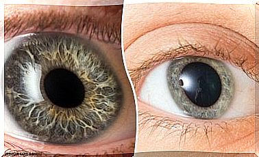
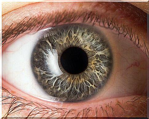
The pupil is that black circle that is seen in the center of the eye. Under normal conditions, the pupil is enlarged when there is little light and it contracts when there is much. Well, anisocoria occurs when the size of the pupil of one eye grows or decreases more than that of the other eye.
The word anisocoria comes from two Greek roots and a Latin one. Thus, it is composed of:
- The Greek prefix “aniso”, which means “inequality”.
- The Greek root “Kore”, which means “pupil”.
- The Latin suffix “ia”, which is “pathology” or “disease.”
So from the etymological point of view, anisocoria is “pathology due to inequality in the pupil “.
It is estimated that at least one in five people has a slight difference between the size of the pupil of one eye and the other. This difference almost always does not exceed half a millimeter. This is called physiological anisocoria.
In these cases it is a normal condition, without any effect on health. However, there are other cases in which this difference in size is a symptom of more complex difficulties.
Causes of anisocoria

Sometimes anisocoria is present from birth. If more than one person in the family has this condition, it is possible to speak of a genetic trait. This is not a red flag.
Sometimes the size of the pupils also changes and then returns to normal. It is unknown why this happens, but it is known to be harmless.
If, on the other hand, the difference in size in the pupils appears suddenly, is more than a millimeter and remains constant, it is possible that there are more serious problems. There may be an eye, vascular , brain, or neurological disease.
The most frequent causes of pathological anisocoria are:
- Brain aneurysm
- Brain tumor or abscess
- Glaucoma
- Internal bleeding in the skull from trauma
- Intracranial hemorrhage, brain swelling, or stroke
- Encephalitis or meningitis
- Seizures
- Lymph node tumor
- Oculomotor nerve palsy
- Migraine
Anisocoria can also be the result of a viral infection, particularly syphilis. There is also a benign condition that affects the size of the pupil and is called Adie syndrome. Likewise, the difference between the two pupils may be due to recent surgery in the area.
Symptoms
Anisocoria is not always easy to detect. Many times it is only discovered accidentally when two photographs are compared and a strange change in the appearance of the gaze is observed.
In fact, anisocoria is considered a symptom, not a disease in itself.
Thus, the assessment of anisocoria depends on the accompanying symptoms.
If it originates from an eye disorder, there is usually also a droopy eyelid, pain, and reduced eye movement. Additionally, you may also experience a headache, fever, and decreased sweating.
The history and medical history are essential to make a correct interpretation of anisocoria. For example, if it appears suddenly, it could speak of a hemorrhagic process in the brain.
If its appearance is gradual and is accompanied by headache, explosive vomiting and double vision, it could suggest the existence of a tumor.
Likewise, when it presents with fever and stiff neck, meningitis may be suspected.
Diagnosis and prognosis

Anisocoria is usually diagnosed by an eye exam. This is done by an ophthalmologist.
Typically, you explore the size of the pupils in a light environment and then in a dark one. This way, you can determine if any of the pupils react abnormally.
If there is a greater difference in size when everything is illuminated, it is concluded that the large pupil is the one that has an abnormal function. If, on the contrary, the sizes differ more when everything is dark, it is concluded that the deficiency is in the smallest pupil.
The goal of the test is to determine if anisocoria is a normal condition or if there are health risks. For this reason, the ophthalmological examination is generally accompanied by an interview.
In this, the moment in which the anomaly was detected is investigated. Questions will also be asked to see if there are other visual or other symptoms.
Depending on the answers, blood or cerebrospinal fluid tests may be ordered. CT scans, MRIs, neck x-rays, tonometry, or others can also be ordered. The prognosis depends on the cause that is generating the anisocoria.