How To Diagnose Heart Disease?
When diagnosing heart disease, doctors confront the patient with information that must be processed and that involves changes in lifestyles. Beyond the medications that exist for an arrhythmia, for example, both diet and physical exercise are tools for controlling heart disease.
A cardiac pathology is one that is located in the heart, unlike cardiovascular diseases that also include the arteries and veins. Examples, in addition to arrhythmias, are dilation of the heart muscle, heart attacks and failure. As for the internal valves, we can mention stenoses and prolapses.
The same complementary methods are used for most of these disorders. Diagnosing a heart disease, in general, consists of following some protocol steps that involve carrying out studies from less to greater complexity. Here we tell you.
What tests can heart disease be diagnosed with?
As we have already mentioned, there are complementary methods to diagnose heart diseases that are common to various pathologies. With an electrocardiogram, for example, it is possible to detect an arrhythmia and a heart attack, without the pathologies being the same.
Electrocardiogram
An electrocardiogram (ECG) is an electrical recording of the activity of the heart muscle. Through electrodes located on the outside of the body, the variations of electricity that are typical of the heartbeat are perceived. This originates from the internal system that the heart tissue has to control the act of beating.
The test does not require anesthesia or extensive preparations. What’s more, it is performed in outpatient clinics and the patient leaves after no more than half an hour of having been there. Sometimes they are routinely requested to track changes that could be hidden, and other times they are requests for subsequent controls on already established pathologies, such as a heart attack or high blood pressure.
The record is captured, through the electrocardiogram apparatus, on a paper with a line that denotes the heartbeat. The interpretation of the study depends on the training of the doctor. There are already established reading protocols that indicate the meaning of each line that was drawn.

Echocardiogram
An echocardiogram may be ordered to diagnose heart disease. The test consists of an ultrasound similar to the one used to monitor a pregnancy, but directed to the heart.
A device called a transducer sends signals, such as echoes, that bounce off the heart muscle and return to be interpreted as an image on a monitor. It is possible to see, live and direct, the movement, the heartbeat, the form and the cardiovascular dynamics.
A variant is the Doppler , with the addition of colors to the image to distinguish between venous blood and arterial blood. At present, it is almost always specified with this modality because the information is greater.
Stress test
The technical name for the stress test to diagnose heart disease is “ergometry.” In basic terms, the heart is stimulated through physical exercise to record what happens under exertional conditions. At the same time, electrocardiograms and echocardiograms will be performed, while the patient is running on a treadmill or pedaling on a stationary bicycle.
There are parameters that are considered in a stress test so as not to exceed capacities that could put the lives of the people who are doing it at risk. Limits have been stipulated for the heart rate, for the incline of the treadmill in case a mobile is used and signs to pay attention to to stop the effort.
It is a very useful complementary method because it simulates real-life situations, in which patients may be forced to run or jog against resistance, as well as climb stairs. In a controlled environment, the heart is studied while replicating reality.
Holter monitoring
The test that doctors shortly call a Holter test is actually a long-duration EKG. Using a device, which patients use for a day or two, the electrical activity of the heart is recorded.
Then, with computer programs, variables such as the numbers of arrhythmias, accelerations, tachycardias, and bradycardias are counted. Its advantage is that there are situations that escape the office ECG and that happen, for example, when the patient sleeps. This is when it is possible to diagnose heart disease that is hidden.
Cardiac catheterization
A cardiac catheterization is a surgical procedure, as it involves inserting a catheter into the circulatory system. Access is usually through the upper or lower extremities, until reaching the heart with the device, which can perform measurements or inject radiographic dyes.
In general, the procedure is supported with external images that will follow the appearance of the radiographic dye. In this way, it is possible to view the heart chambers in special detail that detects anatomical and malfunctions.
Not all patients can undergo catheterization, but those who do have an indication can benefit from treatment at the same time. With the catheter already inserted, it is possible to carry out repairs or remove blockages of clots in the coronary arteries, for example.
Cardiac MRI
Advances in imaging methods have led to a specific resonance imaging (MRI) for the heart. On the same basis as MRIs for the rest of the body, in this case a non-radiant force is used to view the heart muscle. Its indications may overlap with those of the echocardiogram.
CT scan of the heart
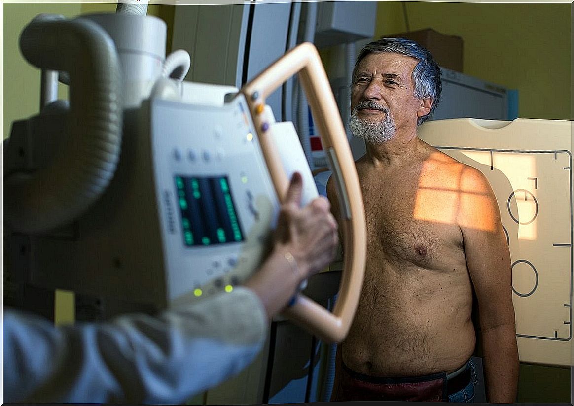
Computerized axial tomography (CT) also responds to the fundamentals of body CT, only applied to cardiac tissue. It uses X-ray radiation, such as radiography, to generate images of the chest that provide more information. It is performed with the patient lying on a stretcher, which is inserted into a tube through which the X-rays will be emitted.
Reduce the risk of heart disease
Beyond diagnosing heart disease, it’s important to prevent it. Current methods allow us to detect life-threatening alterations early, but a proper diet, exercise, stress reduction, and regular check-ups are the best tools.
- Regarding diet, it is convenient to remember that the consumption of vegetables over meats, and the inclusion of natural foods, not ultra-processed, abundant in omega 3, have been shown to protect cardiovascular health.
- Regarding physical exercise, scientific studies agree that aerobic sport practiced on intermediate days, lasting between 30 to 60 minutes, is enough to improve risk.
- To reduce stress there are various techniques that range from deep breathing to yoga, as well as meditation and mindfulness . Each person has an affinity for one or the other according to their personality and their cultural context.
- Finally, regular checks with medical professionals are imposed, at least once a year. An annual electrocardiogram is recommended for adults, although in diabetic people and athletes with a higher range it should be done less frequently, than 6 months.
If all this fails, then it will be necessary to perform a complementary method of those that we list to arrive at the proper diagnosis. When in doubt, it is best to consult a doctor, especially if there are warning symptoms.